


You roll over at 2 a.m. and a deep ache in your right shoulder wakes you up. By breakfast it has faded enough to ignore, until you reach for a seatbelt or hook a bra strap and it returns. Weeks later an X-ray report appears in your patient portal with the phrase "mild degenerative changes," and now you're holding two things that don't seem to fit together. Something is worn, but nobody has explained what that means for your week, your sleep, or your Saturday.
Shoulder osteoarthritis is common, slow, and widely misunderstood, partly because the word arthritis gets used for several different diseases and partly because the scan and the symptom often tell different stories. Knowing what is wearing down, which of the two shoulder joints is involved, and what the evidence does and doesn't support will make your next appointment far more useful than a search engine spiral at midnight.
Table of Contents
- What Shoulder Osteoarthritis Actually Is
- Which Shoulder Joints Wear Down
- Causes, Risk Factors and Who Gets It
- Symptoms That Point to Shoulder Arthritis
- How Doctors Confirm the Diagnosis
- Treatment Options From Rest to Surgery
- Deciding When to See a Shoulder Specialist
What Shoulder Osteoarthritis Actually Is
What is osteoarthritis of the shoulder? It's a degenerative disease of the shoulder joint in which the cartilage that caps the ends of the bones wears thinner over time, and the bone, joint lining and soft tissues around it change in response. Cartilage is the body's low-friction bearing surface, smoother than wet ice, and because it has almost no blood supply it doesn't repair itself the way skin does.
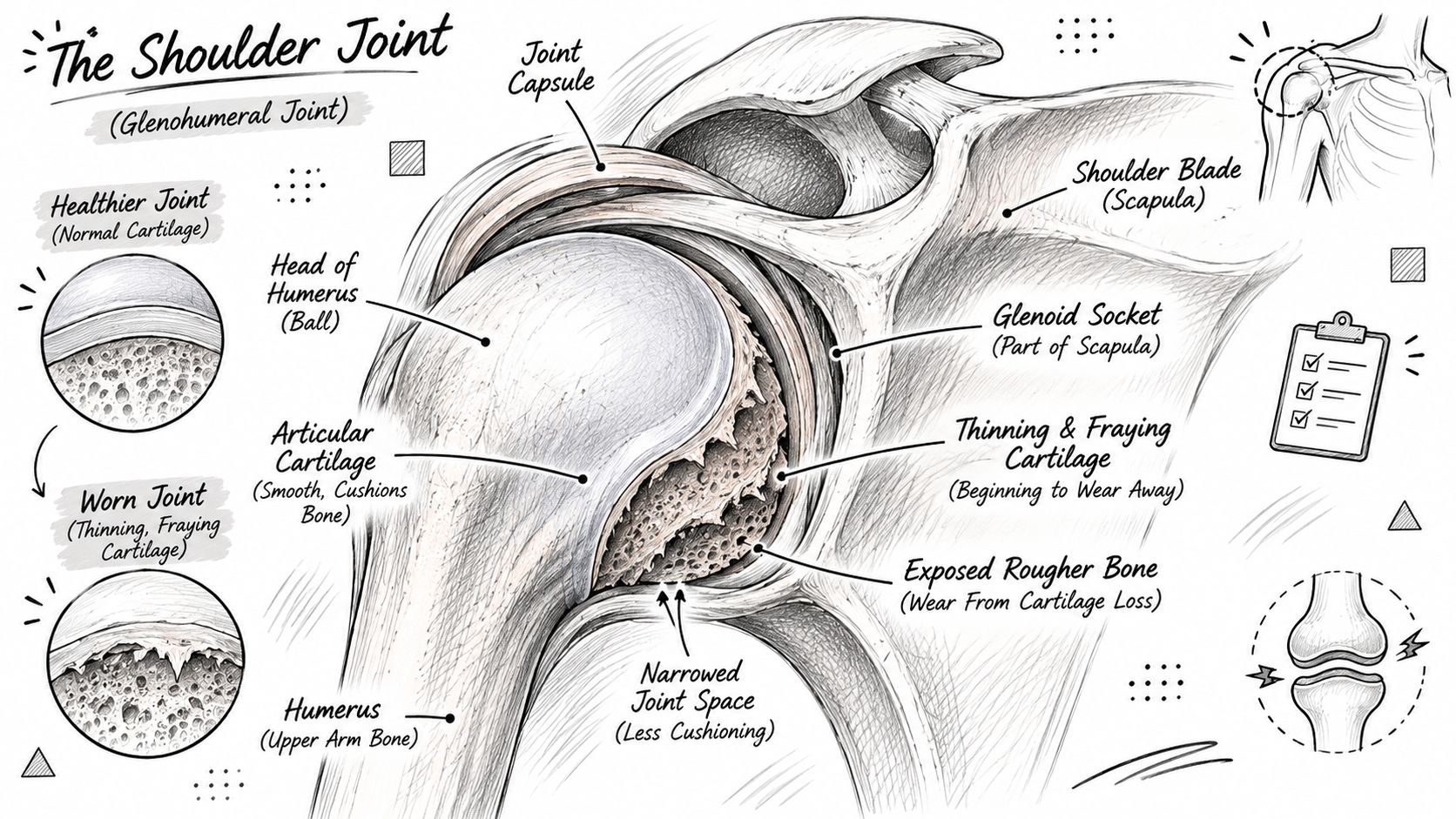
Think about a well-made kitchen drawer. It glides because plastic runners keep metal off metal. Let the runner wear through and the drawer still opens, but it catches, grinds, and eventually gouges the frame. Cartilage does that same job in the shoulder, and the grind you feel is the joint losing its glide.
What changes inside the joint
As cartilage thins, the gap between the bones narrows. The bone directly beneath the worn cartilage hardens, a change called subchondral sclerosis, and the body grows extra bone at the joint edges, which are the osteophytes or bone spurs your report mentions. Small fluid-filled cysts can form in the bone as well.
These changes accumulate over years, not days. That's why patients often describe a gradual stiffening long before they describe sharp pain, and why a single weekend of gardening rarely explains a shoulder that has been tightening since last winter. A past fracture, dislocation or cuff tear can speed the process up, but most cases have no single triggering event. Age is the background factor. And this is not the same disease as rheumatoid arthritis, where the immune system attacks the joint lining from the inside and erodes bone in a different pattern.
Say you reach for a jacket on a hook and feel a deep, grinding ache that wasn't there last year. That's the classic presentation: a mechanical symptom in a joint that has been changing.
A useful test: can your shoulder be described as slow wear over months or years, rather than one sudden tear, pop or fall? Slow wear points toward osteoarthritis. A single event points toward something else, and both can exist at once.
Which Shoulder Joints Wear Down
The shoulder isn't one joint. Two of them can develop osteoarthritis, and where your pain sits is the first clue a clinician uses to tell them apart.
| Joint | Location | Typical pain pattern | Effect on motion |
|---|---|---|---|
| Glenohumeral joint | Ball-and-socket between the upper-arm bone and shoulder blade | Deep ache at the front or back of the shoulder, often spreading into the upper arm | Progressive stiffness and loss of rotation, especially reaching behind the back |
| Acromioclavicular (AC) joint | Top of the shoulder where the collarbone meets the shoulder blade | Localized pain and tenderness at the top of the shoulder | Usually more localized; overall range of motion is often less affected |
The main joint, the ball and socket
The glenohumeral joint is where the rounded head of the upper-arm bone (the humerus) sits against a shallow socket on the shoulder blade (the scapula). It's the joint that gives you the big sweeping motions: reaching overhead, throwing, combing your hair, and rotating your arm behind your back.
Because it carries that range, glenohumeral wear is the version that changes daily life. Losing internal rotation sounds abstract until you can't fasten a bra, tuck in a shirt, or reach the back pocket of your trousers. This joint is also the one most often described as "arthritis of the shoulder" in the first place.
The small joint on top
The acromioclavicular joint sits at the very top of the shoulder, where the collarbone meets the shoulder blade. You can usually find it by running a finger along the collarbone toward the shoulder until you hit a small bump.
Arthritis here tends to be more localized. People point to it with one finger, and it often hurts when they reach across their body or lie on that side. Overall range of motion is frequently better preserved than with glenohumeral disease.
Both joints can be affected at the same time, which is one reason a vague "my whole shoulder hurts" description is less useful than a pointed one. Before your appointment, notice whether the worst of it sits deep in the joint or right at the top, and whether it spreads down the arm.
Causes, Risk Factors and Who Gets It
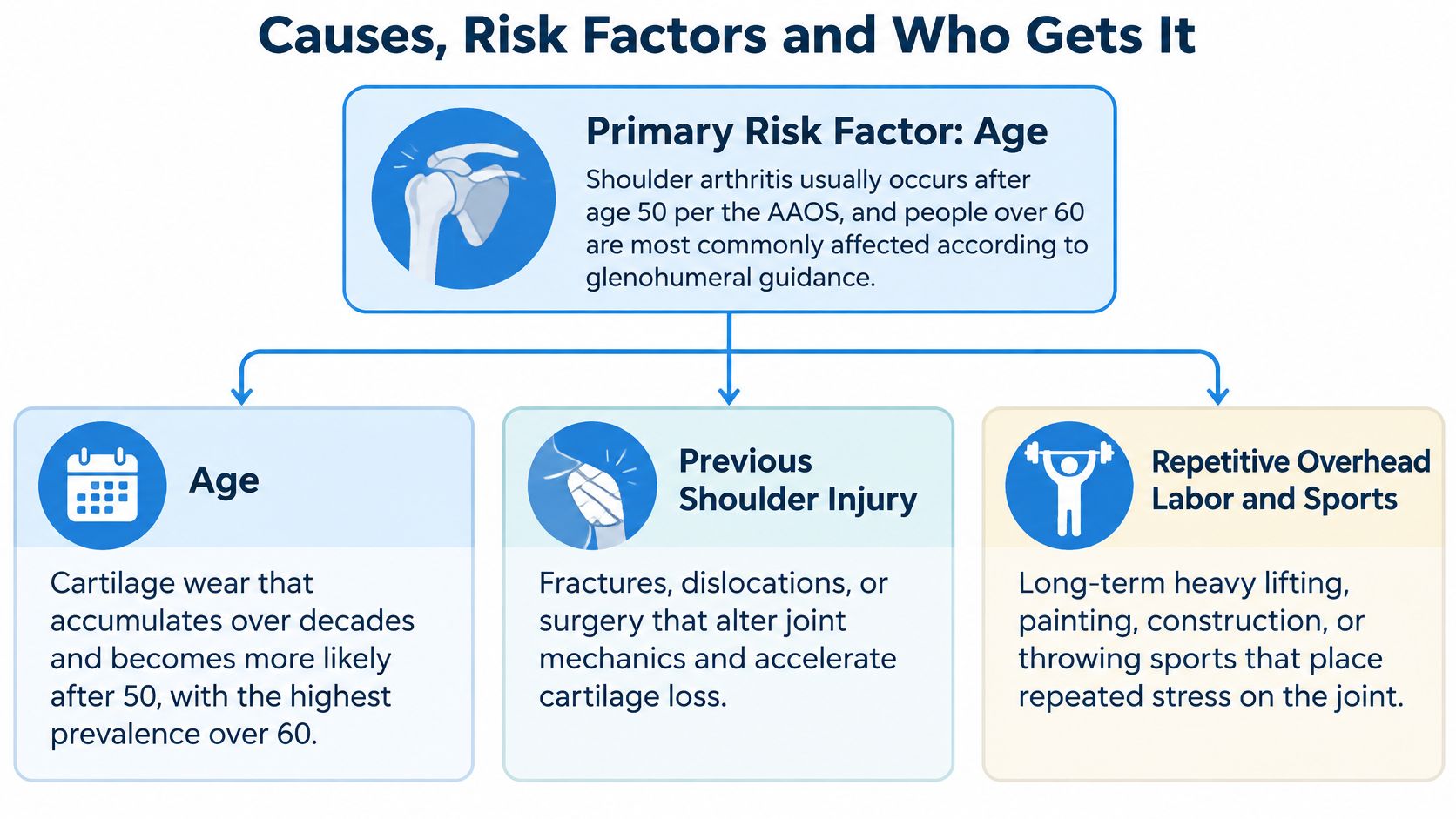
Age is the clearest foundational risk factor. The American Academy of Orthopaedic Surgeons guidance on glenohumeral osteoarthritis states that shoulder arthritis usually occurs after age 50 and identifies people over 60 as the most commonly affected group. In a 2019 primary-care radiograph study of 1,376 patients with shoulder pain, glenohumeral osteoarthritis appeared in 21.2% of cases, with nearly identical rates in women (21.4%) and men (21.0%) and the highest rates among people aged 80 to 89. A separate community study of 541 adults older than 40 found shoulder osteoarthritis in 17.4% overall, rising from 11.1% among those younger than 65 to 20.3% among those 65 and older.
Two things stand out in those figures. Imaging-defined disease is common, and it doesn't favor one sex. Anyone can develop it, and the odds climb with each decade.
What raises the odds without guaranteeing anything
Previous shoulder injury. A fracture, a dislocation, or surgery that changes how the joint surfaces meet can accelerate cartilage loss on that side.
Long-term overhead load. Years of painting ceilings, installing drywall, stocking high shelves, or throwing a ball repeatedly place stress on the same surfaces. Heavy work is a load, not a verdict.
Rotator-cuff disease. The cuff guides how the ball moves in the socket. When it weakens or tears, the joint loads differently and secondary wear can follow.
Inflammatory arthritis. Conditions such as rheumatoid arthritis damage the joint through a separate mechanism. If you're younger and your shoulder is inflamed, hot and stiff in the morning, that's a different conversation. Arthritis that starts early in life usually traces back to injury, surgery or an inflammatory condition rather than decades of use.
A risk factor raises the likelihood. It doesn't schedule the outcome. Plenty of people in their seventies with physically demanding careers have clean joint spaces, and plenty of people who never played a sport develop changes in their fifties.
Here's the joint in motion, which helps make sense of why one surface wears faster than the other.
Symptoms That Point to Shoulder Arthritis
The ache usually sits deep inside the joint rather than on the surface, sometimes felt more toward the back of the shoulder, and it often spreads a few inches down the outer upper arm. It's a dull, heavy soreness rather than a sharp pinch, though sharp catches happen when you move suddenly.
Watch for these patterns, which tend to build gradually.
- Lost rotation behind your back. Fastening a bra, reaching a back pocket, or tucking in a shirt becomes awkward, then difficult, then something you avoid.
- Overhead work gets negotiation. Putting away a suitcase, washing your hair, or painting a wall starts requiring a workaround.
- Crepitus. A grinding or clicking sensation you can hear or feel when the arm moves.
- Night pain. Lying on the affected side wakes you, and sleep becomes the reason you finally book an appointment.
- Morning stiffness. The shoulder feels tight and takes a while to loosen, especially after rest.
Why the X-ray and the pain don't always agree
Visible joint-space narrowing and bone spurs are common findings in middle-aged and older adults, but those structural changes correlate weakly with pain and disability. Many people with clearly abnormal X-rays have few or no symptoms, as Radiopaedia's overview of shoulder osteoarthritis explains. Diagnosis combines your symptom pattern, the physical examination, measured range of motion and imaging, in that order of importance.
Which means an X-ray is one input, not a verdict. A worn joint that doesn't hurt much doesn't need fixing. A painful shoulder with a modest scan needs a search for the actual source.
Several conditions mimic this one closely. Rotator-cuff disease, adhesive capsulitis (frozen shoulder), bursitis, arthritis at the top of the shoulder, and pain referred from the neck nerves can all produce a similar ache. Cuff problems in particular overlap heavily, and what rotator-cuff pain and injuries actually feel like will help you tell a weakness-and-lifting problem from a stiffness-and-grinding problem.
Two questions worth answering honestly: If my X-ray shows arthritis but my pain is mild, do I need treatment? Usually not beyond activity adjustment. And if my pain is severe but the scan looks modest, what else should be investigated? That's when the exam, the neck and the cuff matter more than the film.
Night pain, new weakness, a sudden loss of function, fever, a recent injury, or symptoms that worsen quickly all call for clinical assessment rather than self-treatment. Bring those specifics to a clinician instead of guessing.
How Doctors Confirm the Diagnosis
Diagnosis is clinicoradiographic, which means the history and exam come first and the image confirms what the clinician already suspects. Follow the sequence in order and you'll see why.
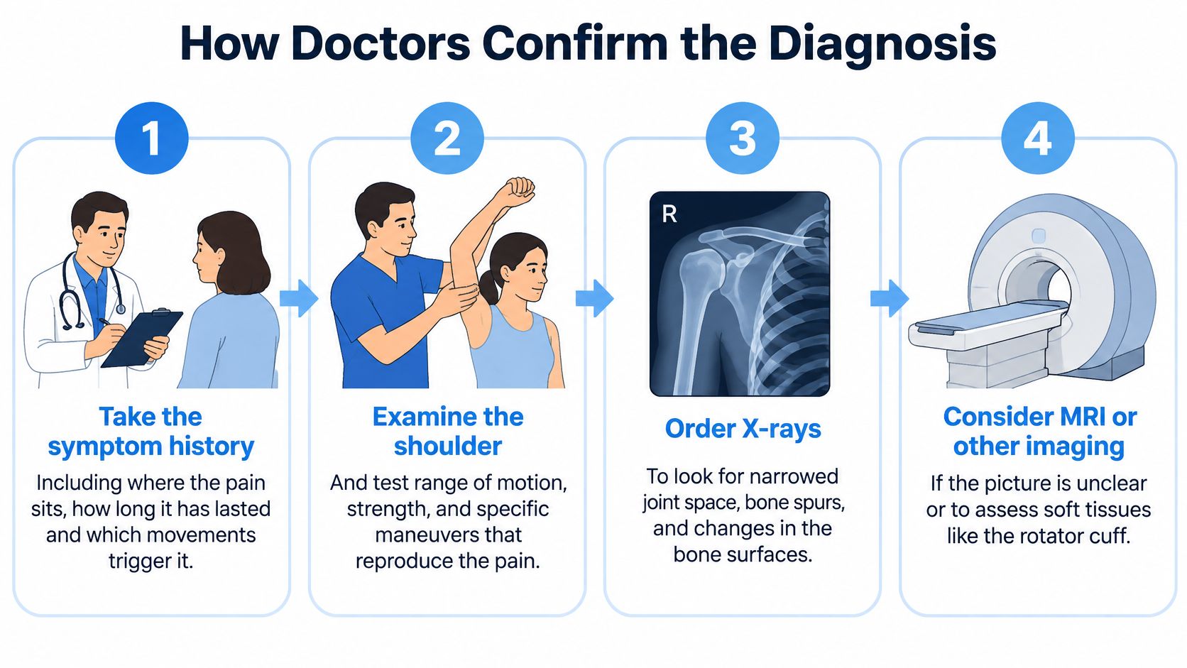
- Take the history. Where exactly is the pain, how long has it been there, which movements trigger it, and does it wake you at night? This step separates the two joints and flags whether the neck is involved.
- Examine the shoulder and neck. A clinician measures active range of motion (what you can do yourself), passive range of motion (what happens when they move it for you), and strength. Passive motion that far exceeds active motion points toward a cuff problem; a hard block in every direction suggests frozen shoulder.
- Order plain X-rays. Standard views are the anteroposterior and axillary films, with a true AP (Grashey) view added to see the joint space with less bony overlap.
- Add MRI, ultrasound or CT only when a specific question remains. These aren't automatic, and they should answer something the X-ray and exam couldn't.
The AAOS plain language summary on glenohumeral osteoarthritis describes the typical X-ray findings: asymmetric joint-space narrowing, inferior humeral-head osteophytes, subchondral sclerosis or cysts, and glenoid bone loss or altered version. Those words are usually what generates the "degenerative changes" line in your report.
| Test | What it shows | When it is used |
|---|---|---|
| Standard X-ray (AP and axillary views) | Joint-space narrowing, bone spurs, subchondral sclerosis | First-line imaging for most patients |
| True AP (Grashey) X-ray | Joint space with less overlap, for clearer assessment of narrowing | Added to standard views to assess joint space |
| MRI | Tendons, soft tissue and cartilage detail | When rotator-cuff integrity is uncertain |
| Ultrasound | Tendons and soft tissue in real time | When tendon integrity is uncertain and MRI is not chosen |
| CT scan | Detailed glenoid bone shape and bone stock | When arthroplasty is being considered |
MRI or ultrasound earns its place when tendon integrity is in question. CT gives the detailed look at glenoid shape and bone stock that matters when replacement surgery is on the table, because implant planning depends on it.
Before agreeing to any scan, ask two things: what question will this answer, and will the result change what we do next? If nobody can answer both, the scan can wait.
Treatment Options From Rest to Surgery
Most plans start with what you can do this week rather than what a surgeon could do next year. The realistic goals are unglamorous and worth stating plainly: keep the motion you have, build strength you can use, take load off the positions that hurt, and protect your sleep.
| Option | Typical use | What to expect |
|---|---|---|
| Activity modification and low-impact exercise | Early or mild symptoms | Helps maintain motion and reduces aggravating loads |
| Supervised physical therapy | Range of motion and strengthening | Benefit specifically for this condition is still a research gap |
| Clinician-directed pain or anti-inflammatory medication | Pain flares that limit daily activity | Short-term pain relief, with dosing set by a clinician |
| Intra-articular corticosteroid injection | Selected patients with substantial pain or disability | Short-term pain and function relief, often time-limited |
| Anatomic total shoulder arthroplasty | Pain and disability persisting despite nonsurgical care, with a functional rotator cuff | Replaces both the humeral head and the glenoid surface |
| Reverse arthroplasty | Significant rotator-cuff dysfunction or severe glenoid deformity | Reverses the ball and socket arrangement to restore function |
Where the evidence gets thin
Physical therapy is the backbone of nonsurgical care, yet its effectiveness specifically for glenohumeral osteoarthritis remains an identified research gap, as the 2025 shoulder osteoarthritis research agenda in the Orthopaedic Journal of Sports Medicine notes. The same AAOS guideline reports insufficient evidence to recommend for or against acupuncture, CBD, capsaicin, glucosamine, chondroitin, cupping and TENS.
Read that carefully. Insufficient evidence isn't the same as proof it fails. It means nobody has run the trials that would settle it, so nobody can promise you a result. What matters is not confusing relief with repair. Feeling better after a session doesn't mean cartilage grew back, because cartilage doesn't regrow on demand.
The honest framing: a treatment can be reasonable to try and still be unproven. Ask what it's expected to change, over what timeframe, and what you'd stop doing if it doesn't help.
Injections often buy a window of relief that lets you move and sleep better, but the window usually closes. Platelet-rich plasma remains investigational for this condition. For day-to-day comfort between sessions, non-compression garments are an option for people who can't tolerate tight sleeves; our Thermo Lite Recovery Dual Shoulder Sleeve, at $74.99, is built for warmth and light daily support, not for changing the joint. And how shoulder heat wraps fit into a daily routine covers the practical side of that.
When surgery enters the conversation
The decision rests on the exam, the imaging, your age, your activity demands and the state of your rotator cuff, not on an X-ray alone. Anatomic total shoulder arthroplasty generally requires a sufficiently functional cuff. Reverse arthroplasty may be chosen when significant cuff dysfunction or severe glenoid deformity is present.
Replacement is also not a novelty. The University of Washington orthopaedics guide to managing glenohumeral arthritis records that prosthetic shoulder replacement dates to 1893, when French surgeon Jules-Émile Péan performed one of the first recorded procedures. In a U.S. national analysis covering 2000 to 2010, the population rate of shoulder arthroplasty for glenohumeral osteoarthritis rose 3.7-fold, with total shoulder arthroplasty up 5.0-fold and representing 80.3% of arthritis-related shoulder replacements in that dataset by 2010.
Deciding When to See a Shoulder Specialist
Some symptoms don't wait for a convenient week. Get assessed promptly for a sudden loss of function, marked new weakness, fever, a recent injury or fall, or symptoms that worsen quickly over days. Those point away from slow wear and toward something that needs a different look.
For everyone else, the trigger is more ordinary. Persistent pain at rest or at night, despite several months of sensible conservative care, is the point where specialist options deserve a discussion.
How to prepare for the visit
Keep a two-week symptom log. Note where the pain sits, which movements bring it on, how often you wake at night, and what helped or made it worse. Two weeks of notes is worth more than a vague "it's been bad for a while."
Bring your old images. Prior X-rays and scans, ideally on disc or through the portal, save you from repeating tests that already answer the question.
Name your top three goals. Sleeping through the night, dressing without help, and returning to overhead work are all legitimate, and they point toward different plans.
Ask what would change the plan. A clear answer tells you the clinician is treating you rather than the radiograph.
Questions worth writing down: which joint is responsible, does the imaging match my symptoms, what nonsurgical plan is realistic and how long should I give it, and what result would prompt a different approach? Wearable devices and supplements shouldn't replace a diagnosis, and the everyday habits that help osteoarthritis work best alongside a plan you understand.
Book the visit. A primary-care clinician can start the workup and refer you; an orthopedic or sports-medicine specialist is the right call if the shoulder has been limiting you for months. Bring the log, bring the images, and ask your questions out loud.
If you're managing shoulder osteoarthritis between appointments, warmth and light support can make the daily routine easier without pretending to be a treatment. Thermo Recovery Wear makes non-compression recovery sleeves, including a dual shoulder sleeve built from infrared-responsive fabric for at-home comfort and light activity. Visit Thermo Recovery Wear to see the Lite collection and find the fit that suits your shoulder.


